|
Role of human papillomavirus type 18 in a subgroup of prostatic cancer with bone metastases: Its protein E2 contains the osteoprotegerin active site.
TRAN Guy Mong Ky (1, 5), KIRKIACHARIAN Serge (2), CAPRANI Adrien (3, 5), MAURISSON Gilbert (4, 5). 1 University Paris-Sud XI ; correspondence: 31 Av du Bois, Châtenay-Malabry, France. E-mail : mkg_tran@yahoo.fr
INTRODUCTION In Japan, the oncogenic human papillomavirus (HPV) type 18 is found by polymerase chain reaction (PCR) in 80% (12/15 cases) prostatic cancer (PC) with bone metastases versus 0% in controls (Anwar K, 1992) . The oncogenic HPV-18,-16,-33 are frequent in a PC subgroup in Germany [HPV-16 in 10/47(21%)] (Serth J, 1999), Canada [13/27 (48.1%)] (McNicolP.J., 1991), Japan (41%) (Anwar K, 1992), in Michigan (USA) [3/23(13%)] (Sarkar F.H., 1996), Italy [6/8(75%)] (Rotola A., 1992) and in Montpellier, France (HPV-16 in 53%) (Moyret-Lalle C., 1995). In Sweden, high antibody levels against HPV-33 multiplied the risk by 2,3 fold (Adami H.O., 2003). In Finnish men, in a prospective study, the presence of antibodies against HPV-16 was associated with an odds ratio (OR) of 2.58 and antibodies against HPV-18 with a statistically significant odds ratio of 2,88 (Dillner J., 1998). Hisada M. (2000) found also an OR of 2.7 for HPV-16 antibodies in a prospective cohort study. Numerous negative results have been reported, and this very important point was analyzed in detail (for tables, see: Terris M.K., 1997 and Strickler H.D., 1998) : Discrepancies are explained in DISCUSSION below : All positive PCR studies (80%, 75%, 53%, 48.1%, 41%, 21%) have used HPV E6 as primers set, and fresh frozen prostatic tissues (except Anwar K, who used archival tissue) ; whereas negative (0%) and weak results employed HPV consensus L1 and archival formalin-fixed, paraffin-embedded tissues . In seroepidemiological studies, the prospective cohort studies were positive whereas the retrospective case-control studies generally negative. The very low concordance of PC in homozygotic twins, (where concordance was observed only 16 times, whereas discordance is 135 times) (Morganti G, 1956) very strongly favoured an environnemental factor : Viral (papillomavirus, polyomavirus) or toxic (cadmium, tobacco). Epidemiology (Sarradet A., 1997) speaks for a sexual transmission [militaries, multipartenarism, herpes virus type 2 infection (Haid M., 1984), prostitutes frequentation , non use of condoms, gonorrhea, syphilis (Hayes R.B., 2000), marriage, wife with uterus cervical cancer, transmitting her HPV to the husband (Lattimer, 1974)]. LINKS BETWEEN HPV MOLECULAR BIOLOGY AND PROSTATIC CANCER. There are many connections between HPV and PC : The HPV oncogènes E6 and E7 are inhibiting the tumor suppressors p53 and pRb, precisely those altered in PC (Sellers W.R., 2002). HPV is integrating upstream of Myc oncogene, which is amplified in 47% of metastatic PC (Kaltz-Wittmer C., 2001). There is a loss of heterozygoty of Bin-1, an anti-myc tumor suppressor, in about half of PC (Sakamuro D., 1996). We found an Epidermal Growth Factor (EGF) motif in HPV E2 (Tran M.K.G., 1993) and EGF receptor ,which gene is on chromosome 7p, is overexpressed in 33.6% of PC (Inoue K., 1998) ; and Trisomy 7 is correlated with a bad prognosis of PC (Bandyk M.G., 1994). There is a » cross-talk » phenomenom between EGF and androgen receptor, that can be activated in the absence of the ligand (Cullig Z., 1997 ; Hobisch A., 1997). EGF transduction pathway is signalling by Ras, which is frequently mutated in PC (Anwar K, 1992). HPV-16 E5 stimulates EGFR (Pim D., 1992).
OBJECTIVE As we have found a molecular mimicry between breast cancer virus 3’ORF and PTHrP (parathormone-related peptide) which could explain bone metastasis in breast cancer (Tran M.K.G., 2003), similarly we tried to elucidate the HPV-18 role in bone metastases, as HPV-18 was so strongly (80% PC versus 0% controls) (Anwar K, 1992) and specifically associated with the presence of bone metastasis. HPV-18 is known to induce a severe, aggressive cervical cancer in women. HPV-18 has been found also in a metastatic lymph node , and in a seroepidemiological study the OR is 2.88 when antibodies against HPV-18 are present (Dillner J, 1998). To find a molecular biology explanation would strengthen the role of HPV-18 in the occurrence of a subset of particularly aggressive and lethal PC, that resist to hormonal treatment.
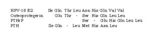
METHOD Comparison of aminoacids (AA) sequences between HPV-18 E2 protein and 3 proteins of calcic homeostasis : Osteoprotegerin (OPG), PTH (parathyroid hormone) and PTHrP (PTH-related protein). The screening sequence is the tetrapeptide HQLL common to osteoprotegerin and PTHrP.
RESULT Osteoprotegerin is the most homologous to HPV-18 E2, spanning 8 aminoacids, which is significant . A contrario, histidine (His) is absent in benign HPVs.
DISCUSSION Owing to the low HPV copy number in prostatic tissue (Serth J., 1999), the PCR seemed the most appropriate method to reveal HPV in prostatic cancer. But the choice of the primer sets is crucial and can alone determine the PCR positivity (Terris M.K., 1997) : In the same patient, E6 primer set obtained 11/53 (20.8%), compared to 0/53 (0%) for L1 primer. Previous numerous negative results have been reported, throwing doubt, and this very important point was analyzed in detail (for review tables, see: Terris M.K., 1997 and Strickler H.D., 1998) : We can now clarify further the debate and try to explain some of these apparent discrepancies, on the basis of 2 criteria (first, the primer sets used, second, the prostatic tissue preparation) as well as the PC heterogeneity. POSITIVE RESULTS: All positive PCR studies (80%, 75%, 53%, 48.1%, 41%, 21%) have used HPV E6 as primers set, and fresh frozen (Zambrano A., 2002) prostatic tissues (except Anwar K, who used paraffin tissue and E6 primer; like Sarkar who obtained only 13%) . NEGATIVE RESULTS: Whereas almost all negative (0%) and weak (about 15%) results employed HPV consensus L1 and archival formalin-fixed, paraffin-embedded tissues ; these » L1-paraffin » authors were numerous : Serfling U (0%), Effert P.J. (0%), Terris M.K. (0%) Tu H. (2.3%), Ibrahim G.K. (16%), Suzuki H. (16%), Widerhoff L. (12.5%) (15.2% in whites). Wideroff L. is the only author to obtain no result with E6 primers set, like Noda T. (1998) [0/38 (0%)] in paraffin tissue. It seems that paraffin is not recommanded for HPV research, because the majority of results are negative or very weak. Iwasawa A (1990) and Gherdovich S (1997) have negative results, but their patient number is statistically too low (n=5 only). Masood S. (1991) had negative results but used in situ hybridization instead of PCR. There is some doubt on the results of Strickler H.D., 1998, who found zero HPV everywhere (cancer and adenoma controls) : There is no explanation for their failure (they correctly used E6 and frozen tissue for PCR), except the PC heterogeneity (they may have examined other PC subsets linked to cadmium, polyomavirus and genetics). It is unknown why Strickler H.D. did not use the same E6 primers set than for example Anwar. A slightly different sequence may be crucial for perturbing the results. For the discrepancies in seroepidemiological studies, which are less convincing than PCR studies, the criteria used for discussion is whether they were prospective or retrospective (retrospective is more subject to errors, because the choice of controls can always be debated) : The prospective cohort studies were positive (Hisado M., 2000 ; Dillner J. 1998), whereas the retrospective case-control studies generally negative (Strickler H.D., 1998 ; Rosenblatt K.A., 2003 ; HayesR.B.,2000 ; Adami H.O., 2003). However, Adami H.O. found negative results with HPV-16 and -18, but positive result with HPV-33 (OR = 2.3). All these HPV positive results are concerning a subset of PC (about 30-40%), but it must be kept in mind that non-HPV linked PC exist, for example PC related to tobacco, cadmium (toxic) or polyomavirus (viral) (Zambrano A., 2002) or genetic. So a large PC heterogeneity can also explain as well negative (Strickler H.D., 1998) or weak results.
CONCLUSION The PC subgroup with bone metastases in Japan is linked by PCR in 80% cases to HPV-18, versus 0% in controls. HPV-18 E2 protein contains an OPG motif. This confirms the viral etiology of a PC subgroup, and explains the mechanism of bone metastases formation. Cidofovir (Vestide ®) is an active anti-viral against HPV (De Clerq E., 2003) and is also used as a 1% gel in cervix intraepithelial neoplasia grade III (Snoeck R., 2000). Imiquimod, a imidazoquinoline immune system stimulator, has been approved for the therapy of genital warts (Garland S.M., 2003). An anti-HPV-16 vaccine succeeded against cervical cancer in a clinical trial (Koutsky L.A., 2002). If a subset of PC has to be considered as a sexually transmitted disease, it would theorically follow the same rules as for every STD, i.e. obligatory declaration, systematic viral (HPV, polyomavirus) research in the partner and simultaneous treatment of the partner to avoid any ping pong reinfestation. Obviously, none of these sanitary measures is effectively entered in routine today. There is also a need to develop good and fiable serological tests, alike those used in women cervical cancer (virus-like particles or VLPs) in a cohort prospective seroepidemiological study, and respect the conditions of a highly sensitive nested PCR with specific E6 (not L1) primers set (Terris M.K.,1997 ; Anwar K, 1992) on fresh frozen (not paraffin-embedded archival) prostatic tissue, without forgetting to research associated polyomavirus (Zambrano A., 2002). The spectrum of oncogenic HPV types being the most exhaustive as possible.
REFERENCES Adami H.O. et al.. Cancer Epidemiol Biomarkers Prev 2003, 12 : 872-5. Bandyk M.G. et al. Genes Chromosomes Cancer 1994, 9/ 19-27. De Clerq E. et al. Clin Microbiol Rev 2003, 16 : 569-96. Effert P.J. et al. J Urol 1992, 148 : 192-4. Garland S.M. Curr Opin Infect Dis 2003, 16 : 85-9. Haid M. et al. Urology 1984, 24 : 623-5. Inoue K et al. Acta Med Okayama 1998, 52 : 305-10. Kaltz-Wittmer C et al. Lab Invest 2001,80 : 1455-64. Lattimer, 1974 Masood S et al. South Med J 1991, 84 : 235-6. Pim D. et al. Oncogene 1992, 7 Rosenblatt K.A. et al. Cancer Epidemiol Biomarkers Prev 2003, 12 : 763-8. Sakamuro D. et al. Nature Genet.1996, 14 : 69-77. Terris M.K. et al. Urology 1997, 50 : 150-6. Wideroff L. et al. Prostate 1996, 28 :117-23. Zambrano A. et al. Prostate 2002, 53 : 263-76.
|
Luttons contre le Sida
©2024 Positifs.org All Rights Reserved. | Réalisation : Beaba-Informatique | Politique de confidentialité | Positifs
Laisser un commentaire